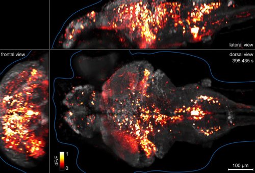
fennetic: Whole-brain functional imaging at cellular resolution using light-sheet microscopy (o
fennetic:Whole-brain functional imaging at cellular resolution using light-sheet microscopy (original video file 20MB)Here we use light-sheet microscopy to record activity, reported through the genetically encoded calcium indicator GCaMP5G, from the entire volume of the brain of the larval zebrafish in vivo at 0.8 Hz, capturing more than 80% of all neurons at single-cell resolution.some background:young zebrafish fry, commonly known in the aquarium trade as danios, are completely transparent. this makes them ideal for neuroscience since we can see everything happening at once. scientists would like to use a voltage sensitive dye, but current dyes are too slow to show individual firing events. so for now we have to genetically engineer fish with built-in reporter proteins, in this case a calcium concentration dependent green fluorescent protein.a specially engineered fish is held in a block of clear gel, the microscope scans a blue laser beam across the fish 33 times a second, and a computer reconstructs the 3D volumetric image about once a second. the firing nerve cells actually glow green, not orange. at 385 seconds the fish sees something in its environment that startles it. this causes an explosion of nerve activity that slowly burns out. after the experiment, the fish was returned to its aquarium and monitored for changes. the scientists then attempted to map out the circuits of its brain and guess at how the neurons talk to each other. questions for the class:why did the scientists use a blue laser, wouldn’t the flickering lights give the fish a seizure?why use a laser at all? why not just rotate the fish or the camera?there are no hypotheses, experiments, or conclusions. is this science? -- source link
Tumblr Blog : fennetic.tumblr.com
#science


