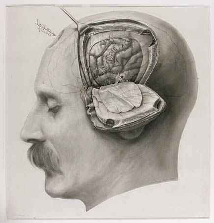
photograph/drawing/diagram of the human head that had undergone craniectomy. Revealing the temp
photograph/drawing/diagram of the human head that had undergone craniectomy. Revealing the temporal lobe. moving outward we see the meninges tissue which is a connective tissue that holds the brain in place. next on our way out, we see the skull that was cut out using a larger more primitive and risky cranial drill. todays cranial drills offer small drill bits to decrease the risk factor of harming the patient. but it looks like this one is already dead. moving on out we see the skin that is connected by more fibrous tissues. further explanation is given below. the CNS is surrounded by connective tissue membranes, called meninges, and by cerebrospinal fluid. There are three layers of meninges around the brain and spinal cord. The outer layer, the dura mater, is tough white fibrous connective tissue. The middle layer of meninges is arachnoid, which resembles a cobweb in appearance, is a thin layer with numerous thread like strands that attach it to the innermost layer. The space under the arachnoid, the subarachnoid space, is filled with cerebrospinal fluid and contains blood vessels. The pia mater is the innermost layer of meninges. This thin, delicate membrane is tightly bound to the surface of the brain and spinal cord and cannot be dissected away without damaging the surface. -- source link
Tumblr Blog : bodyparks.tumblr.com
#diagram#tissue#historical#medical terminology
