neuromorphogenesis:After Death, H.M.’s Brain Uploaded to the CloudThe most famous neuroscience patie
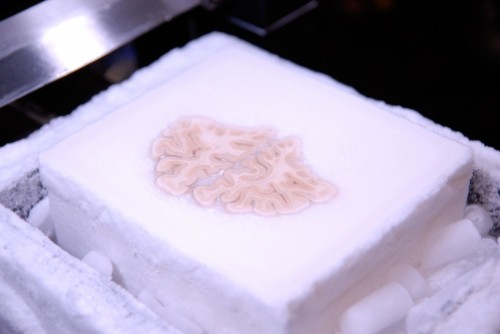
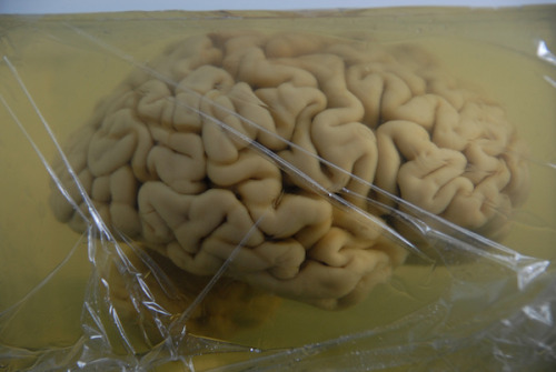
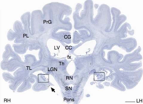
neuromorphogenesis:After Death, H.M.’s Brain Uploaded to the CloudThe most famous neuroscience patient of the modern era died of respiratory failure on December 2, 2008, at 5:05 PM Eastern Time. Not long after, the body of 82-year-old Henry Molaison — known simply as H.M. in hundreds of scientific papers — went on a two-hour ride from his nursing home in Windsor Locks, Connecticut, to Massachusetts General Hospital in Boston. The cadaver spent the next nine hours inside of an MRI machine, getting scanned every which way.Meanwhile, on the other side of the country, neuroanatomist Jacopo Annese rushed to the San Diego airport and boarded a red-eye flight to Boston. After landing, and pounding a few cups of coffee, he went to the hospital to join neuropathologist Matthew Frosch in the meticulous and high-stakes extraction of H.M.’s brain.H.M. had one of the most important brains in the world, at least if you ask a neuroscientist. In 1953, at age 27, he underwent experimental brain surgery to treat the terrible seizures that had plagued him since childhood. The seizures quieted after surgeon William Beecher Scoville removed pieces of the temporal lobes above his ears — including, notably, large parts of the hippocampus — but it came at the cost of permanent amnesia. For the rest of his life, H.M. could only hold on to memories of events that happened before his surgery.Though he couldn’t remember what he had for breakfast, H.M. could learn new motor memory tasks and had normal intelligence, illustrating both the specificity of the hippocampus and the multifaceted nature of memory. All of this we know thanks to decades of work by Suzanne Corkin and her colleagues at McGill University and MIT. As Corkin writes in her fascinating new book Permanent Present Tense*, “Henry’s disability, a tremendous cost to him and his family, became science’s gain.”H.M. not only participated in hundreds of studies while he was alive, but donated his brain to science. The morning after H.M. died, a groggy and jet-lagged Annese knew that this brain tissue might pay scientific dividends for many decades to come — but only if the extraction went smoothly.“I had these nightmares about Young Frankenstein — you know, when he drops the brain?” Annese recalls, laughing. “This artifact is so important, and you can create big damage when you’re doing an autopsy if you’re not careful.” Luckily the scientists recovered the brain without making any nicks. So far, so good, Annese told himself.H.M.’s brain stayed in Boston, suspended upsidedown in a standard formaldehyde buffer, until February. Then Annese took it to California, to his lab at The Brain Observatory at the University of California, San Diego. Over the next 10 months, Annese gradually added sucrose to the buffer, creating a cryoprotectant that would allow the entire brain to be frozen without forming tissue-damaging ice crystals.On the one-year anniversary of H.M.’s death, Annese’s team froze his entire brain as a single block and began a 53-hour process of cutting it into some 2,400 super-thin slices. “I didn’t sleep for three days,” Annese says. He had a team of students that took shifts to help him — and to make sure he stayed awake. ”There was always one person next to me, and if I looked like I was phasing out or missing a slice, the code word was ‘prosciutto’,” says Annese, who is Italian. “It was probably the most engaging, most exciting thing I’ve ever done.”Annese’s team live-streamed and Tweeted the entire procedure, and he says some 400,000 people tuned in to watch. In the end, just two slices were damaged while cutting.A camera mounted above the iced brain took a high-resolution picture of each slice. These images became the basis of a digital, interactive atlas of H.M.’s brain, as Annese and Corkin describe in Nature Communications. The atlas will be made available to any other researcher interested in collaboration, Annese says.On image 3 the boxes on the picture, you are looking at what was left of H.M.’s hippocampus. Although most emphasis on H.M. has focused on his missing hippocampus, brain-imaging studies have shown since the late 1990s that he actually retained about 50 percent of this region. The new resource confirms this, and also allows researchers to look closely at individual hippocampal cells, providing a fine-grained molecular view that is not possible with brain imaging.“Imaging can only go so far,” says David Amaral, director of research at the M.I.N.D. Institute at the University of California, Davis, who worked on a 1997 brain-imaging study of H.M.’s brain. “We knew that there was a portion of the hippocampus that was intact. What you can’t see in MRI is whether the neurons are there and what state they’re in. Now that’s come out in this new paper.”Those hippocampal neurons look fairly normal, which comes as a surprise to Menno Witter, a memory expert at the Kavli Institute for Systems Neuroscience in Trondheim, Norway, who was not involved in the work. That’s because H.M.’s surgery removed not only parts of the hippocampus, but the entire ‘entorhinal cortex,’ a region that connects the hippocampus to the cortex, or outer layers of the brain. In animal models, Witter says, “removal of entorhinal inputs generally lead to striking changes” in the hippocampus. (It could be that these changes did happen in H.M.’s brain, but won’t be obvious until researchers compare it with more control samples, he adds.)Even without inputs from the entorhinal cortex, it’s possible that H.M.’s remaining hippocampus was partially connected to the brain stem and other, non-cortical areas of the brain. So it could have had a functional role in H.M.’s cognition, Witter says. “What that is, I would find that hard to predict.”More generally, this new paper and other research on H.M. over the years has gradually shifted from a focus on individual regions, such as the hippocampus, to the interaction of many connected regions. “There is certainly agreement that the hippocampus is critical for episodic memory,” says Rebecca Burwell, a memory researcher at Brown University who was not involved in the work. “But these new findings confirm that understanding brain circuits, in this case the pathways in and out of the hippocampus, is the key to understanding how the brain supports memory and other cognitive processes.”H.M.’s brain is the most famous of about 60 brains that are currently undergoing Annese’s new freezing-and-cutting procedure, and 300 living people have signed up to donate theirs. Annese hopes that his digital approach will become “a radical new way of brain banking,” where brain tissues are saved from dusty storage rooms and uploaded to a more lasting digital warehouse.The most fascinating thing about the H.M., and the new H.M. atlas, has to do with the last decade of his life, in which he developed severe dementia. It could have been Alzheimer’s, and researchers are currently searching H.M.’s brain samples for the disease’s signature amyloid-beta plaques. One theory of Alzheimer’s says that the disease begins in the entorhinal cortex. That idea would be called into question if H.M. indeed had the disease.And even if H.M. didn’t have Alzheimer’s, his dementia still leaves a puzzle for brain researchers. “What will be of interest is what we can make of how intact H.M.’s brain is given that he was undergoing this dementing illness,” Amaral says. “To me, that’s one of the major unanswered questions about Henry. And something else that Henry can teach us.” -- source link
Tumblr Blog : themedicalstate.tumblr.com
#science#neuroscience#hm#history#neuroanatomy#dimentia#case study#neurology


