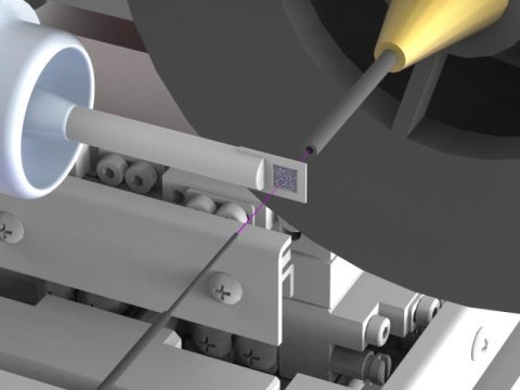
Crystals in a pink X-ray beamSerial crystallography using broad-spectrum X-rays at a synchrotron sou
Crystals in a pink X-ray beamSerial crystallography using broad-spectrum X-rays at a synchrotron sourceA newly developed experimental set-up allows the X-ray structure determination of biomolecules such as proteins with far smaller samples and shorter exposure times than before. At so-called synchrotron sources, protein crystal can be studied considerably more efficiently and quickly by using broad-spectrum X-rays. However, due to the large amount of scattered radiation, this has until now required very large crystals. The newly developed experimental set-up now allows the unwanted scattered radiation to be substantially reduced, so that scientists have been able to perform serial crystallography using broad-spectrum synchrotron radiation for the first time. The international team led by DESY scientist Alke Meents published its findings from experiments at the Advanced Photon Source (APS) in the U.S. in the journal Nature Communications.Synchrotron sources are circular particle accelerators that produce bright X-ray radiation. These X-ray sources are the workhorses for protein structure determination. To elucidate the spatial structure of a particular protein, crystals are grown from it and investigated with X-rays at a synchrotron. The crystal diffracts the X-rays in a characteristic manner, and from the resulting diffraction pattern the inner structure of the crystal, and with it the structure of the protein can be calculated down to the atomic level.Read more. -- source link
Tumblr Blog : materialsscienceandengineering.tumblr.com
#materials science#science#x-rays#synchrotron#biomaterials#proteins#crystals#x-ray crystallography#optics
